
From Fat to Fibrosis: Recognizing MASLD Before It Becomes Advanced Liver Disease
Aug 17, 2026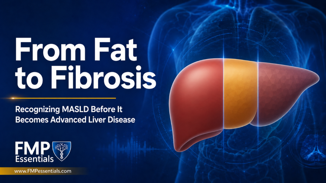
Metabolic dysfunction-associated steatotic liver disease (MASLD) often begins quietly. A patient may feel well and remain unaware that fat is accumulating in the liver.
For many patients, hepatic steatosis remains stable or improves with intervention. For others, it progresses to inflammation, fibrosis, cirrhosis, and potentially liver failure.
In a recent episode of the FMP Essentials Show, Dr. Ellie Campbell and Dr. Yousef Elyaman explore this progression through the story of Dr. Campbell’s close friend, who had polyendocrine metabolic ovarian syndrome (PMOS), formerly called polycystic ovary syndrome (PCOS), and diabetes. Her liver disease was not identified until she already had cirrhosis.
Her story raises an important question: How can practitioners recognize patients at risk before silent liver disease becomes advanced?
Liver Fat and Liver Fibrosis Are Not the Same
Steatosis refers to excess fat stored in the liver. Although important, it does not reveal how much permanent damage has occurred.
When fat accumulation is accompanied by inflammation and cellular injury, the condition may progress to metabolic dysfunction-associated steatohepatitis. Repeated injury can then activate the liver’s wound-healing response, causing scar tissue to accumulate.
This scarring is called fibrosis. Early stages indicate limited scarring, while stage 3 represents advanced fibrosis. Stage 4 is cirrhosis, in which extensive scarring changes the liver’s structure and interferes with its function.
The distinction matters because fibrosis severity is one of the strongest predictors of liver-related complications and mortality in MASLD. Identifying liver fat is useful, but determining whether fibrosis has developed is often more clinically important.
Why Advanced Liver Disease Can Be Missed
The liver can continue performing essential tasks after significant damage has occurred. Patients may therefore remain asymptomatic until disease is advanced.
Even patients with fibrosis may feel relatively well. Fatigue, abdominal discomfort, jaundice, swelling, confusion, or gastrointestinal bleeding may not appear until later stages.
In the case discussed during the podcast, advanced disease became apparent after gastrointestinal bleeding led to the discovery of enlarged veins around the esophagus and stomach, known as varices. By then, cirrhosis and portal hypertension were already present.
What Portal Hypertension Reveals
Blood from the digestive organs normally flows through the liver. When cirrhosis makes the liver stiff and resistant to blood flow, pressure can build within the portal venous system.
This is called portal hypertension.
The backed-up blood may enlarge veins in the esophagus or stomach. These varices can rupture and cause life-threatening bleeding. Portal hypertension may also enlarge the spleen, promote abdominal fluid accumulation, and contribute to a falling platelet count.
A declining platelet count can therefore be more than a hematologic finding. In the appropriate context, it may provide a clue to advancing fibrosis or portal hypertension. Reduced production of thrombopoietin, a liver-derived hormone involved in platelet production, may also contribute.
Platelet changes are not specific to liver disease, but an unexplained downward trend should not be ignored.
Move Beyond Detecting Liver Fat
An abdominal ultrasound may identify hepatic steatosis, but it can miss mild fat accumulation and cannot reliably stage fibrosis.
Several noninvasive tools can help practitioners estimate fibrosis risk:
- The Fibrosis-4 Index uses age, aspartate aminotransferase, alanine aminotransferase, and platelet count.
- The Enhanced Liver Fibrosis test measures biomarkers associated with liver scarring.
- Vibration-controlled transient elastography, commonly known as FibroScan, estimates liver stiffness and hepatic fat.
- Magnetic resonance elastography provides another imaging-based assessment when available.
No single test is perfect. The Fibrosis-4 Index is also less reliable in adults younger than 35, which is relevant when evaluating younger patients with PMOS or early metabolic dysfunction. Results should be interpreted alongside metabolic risks, laboratory trends, imaging, and medical history.
Know When to Involve Hepatology
Patients with evidence of advanced fibrosis, cirrhosis, portal hypertension, or conflicting noninvasive results generally warrant evaluation by a hepatologist.
Referral does not mean the functional or primary care practitioner no longer has a role. Nutrition, physical activity, glucose regulation, alcohol reduction, sleep, and management of associated cardiometabolic conditions remain important.
However, patients with advanced disease may also require specialized monitoring for varices, liver cancer, impaired liver function, and transplant eligibility. The goal is to identify these patients before an emergency reveals the diagnosis.
Intervene Before Fibrosis Becomes Advanced
The earlier MASLD is recognized, the greater the opportunity to address its underlying drivers. Exercise can reduce liver fat even without substantial weight loss. Sustainable dietary changes, improved insulin sensitivity, reduced alcohol exposure, and management of diabetes and other cardiometabolic risks can also support liver health.
Once significant fibrosis develops, management becomes more complex. Practitioners should therefore move beyond asking whether liver fat is present and consider whether the disease has begun to scar the liver.
Clinical Takeaway
MASLD exists along a spectrum from potentially reversible steatosis to inflammation, fibrosis, cirrhosis, and liver failure.
By combining metabolic risk assessment with platelet trends, noninvasive fibrosis testing, appropriate imaging, and timely specialist referral, practitioners can identify higher-risk patients before silent disease becomes a medical emergency.
References
- Rinella ME, Neuschwander-Tetri BA, Siddiqui MS, et al. AASLD Practice Guidance on the Clinical Assessment and Management of Nonalcoholic Fatty Liver Disease. Hepatology. 2023;77(5):1797–1835.
- Tacke F, Horn P, Wai-Sun Wong V, et al. EASL–EASD–EASO Clinical Practice Guidelines on the Management of Metabolic Dysfunction-Associated Steatotic Liver Disease. J Hepatol. 2024;81(3):492–542.
- Manzano-Nunez R, Santana-Domínguez M, Rivera-Esteban J, Pericàs JM. Non-Alcoholic Fatty Liver Disease in Patients with Polycystic Ovary Syndrome: A Systematic Review, Meta-Analysis, and Meta-Regression. J Clin Med. 2023;12(3):856.
- FMP Essentials Show. Dr. Ellie Campbell and Dr. Yousef Elyaman on PMOS and metabolic dysfunction-associated steatotic liver disease.
Author

